




Inside a room on the fourth floor of the Chemistry Annex, a mechanical leviathan helps UC Davis scientists unravel the mysteries of chemical biology. Gray in color and rectangular in shape with a fin-like protrusion atop it, the instrument is affectionately referred to as “Bruce” by UC Davis Assistant Professor of Chemistry Elizabeth Neumann and her colleagues.
Named after the antagonist in the 1975 film Jaws, Bruce is a timsTOF fleX system, a matrix-assisted desorption/ionization mass spectrometer, or MALDI-MS. To showcase the influence of its namesake, the instrument is outfitted with paper teeth and googly eyes, the latter officially stamped by Bruker Scientific with their logo on the pupils.
To understand health and disease development, scientists need a comprehensive picture of the cellular and molecular interactions occurring in the body. Bruce helps Neumann and her colleagues accomplish just that.
Developed by Bruker Scientific with the assistance of Neumann, Bruce enables the high-throughput chemical analysis of single organelles to whole body systems without removing them from their biological and structural context.
The data gathered by Bruce is dense and the instrument can even measure usually hard-to-label molecules such as lipids, glycans and neurotransmitters.
“But I can convert that data into something else,” Neumann said. “An image.”
And those images are spectacular, reminiscent of abstract paintings and stained-glass windows with their kaleidoscopic quality.
“Cells actually interface with one another in neighborhoods and communities and the timsTOF fleX system allows us to do such high-resolution imaging,” said Neumann. “We’re at a spatial resolution of 5 to 10 microns and this gives us an idea of what these neighborhoods start to look like and how they’re functioning.”
Scroll on to check out some of our favorite imagery from the Neumann Lab and learn a bit about the research behind them.
A spinal cord

As a MALDI-MS, Bruce’s imaging capabilities push beyond the soft tissue of the body: organs such as the brain, liver and kidney. In collaboration with graduate student Kayle Bender and Professor Aijun Wang, of the departments of surgery and biomedical engineering, Neumann has extended Bruce’s analytical powers to non-soft tissues of the body such as the spinal cord.
“Everyone said this project was impossible,” said Neumann as she showcased a molecular image of the major components of a spinal column. The cells in the image are colored in yellow (grey matter), blue (white matter), teal (fluid), green (blood vessel), pink (bone/bone marrow) and red (muscle).
With the MALDI-MS, the team (specifically, Wang and his graduate student Arya Lall) is using mouse models to study the development and progression of spina bifida, a condition that detrimentally affects the development of a baby’s brain and spinal cord. The condition can lead to paralysis among other conditions.
Alzheimer’s disease

Alzheimer’s disease is characterized by the buildup of beta-amyloid plaques and excess lipids in the brain. It’s thought that the reduced cognitive function characteristic of diseases like Alzheimer’s can be linked to diet. And it seems that the western diet — one that is high in fat and sugar — is particularly harmful.
To understand the relationship between diet and brain health, Neumann and graduate student Catelynn Shafer have used Bruce to image the brains of mice with Alzheimer’s disease, creating a spatial map of lipid distribution.
In their experiments, they’ve found that mice that were fed keto diets showcased a decrease in lipids of interest in the brain. While the research is still in its early stages, it shows that MALDI-MS is a promising technique for finely tracking how the molecules in our food affect the brain chemistry of those with Alzheimer’s disease.
Gene expression
Graduate student Kyle Vanderschoot, a member of the Neumann Lab, has used Bruce to develop a method to map gene expression in tissue samples. Such spatial transcriptomics are usually prohibitively expensive, sometimes costing tens of thousands of dollars.
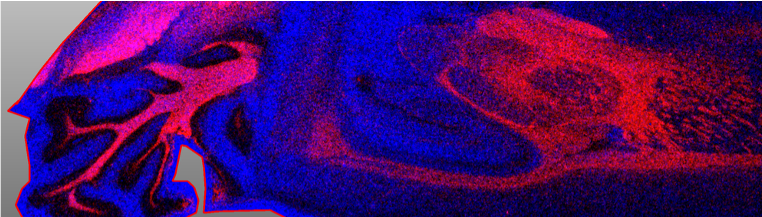
But Vanderschoot developed a technique that first lowered the cost to $500 per probe. His second iteration lowered that price point even further to $15 per probe.
“What this allows us to do is take a pixel and actually say, ‘Here’s the genes expressed by the cell and here’s its metabolism,” said Neumann.
Whole animal system imaging

Bruce has also been harnessed for whole animal system imaging. In one image, a mouse’s internal body is illuminated in a tapestry of neon colors.
Graduate student Karina Vargas, a member of the Neumann Lab, is applying this imaging method to understand how nanoplastics are distributed through the body following ingestion.
“It turns out there are very few mass spectrometry methods for understanding plastic,” said Neumann, noting the dire need for such a technique in today’s environment.
In their experiments, the team followed this distribution process in mouse models that ingested the plastic polystyrene. After imaging, the team found evidence of plastic in the stomachs, intestines, hearts, lungs, livers and brains of their mouse models.
“By visualizing this in the whole animal system, we were able to actually follow and add context to where polystyrene is ending up in each of these organs,” Neumann said.
Ovarian cancer
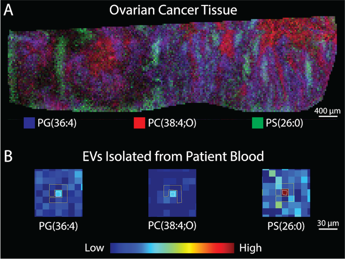
In collaboration with Randy Carney, an associate professor of biomedical engineering, Neumann and graduate student Kayden Lynch are using Bruce’s analyzation powers to study ovarian cancer. Specifically, they’re studying nanoparticles known as extracellular vesicles, or EVs. These molecules can range in size from 30 to 100 nanometers.
“A tumor cell ends up spewing all of these EVs and it’s hypothesized that EVs are involved in things like metastasis,” Neumann said. “What we hypothesize is that the chemical constituents of these EVs probably match the host cell that they come from.”
Neumann and her colleagues have found that EVs and their parent tumor cells share certain chemical signatures.
“What we’re trying to understand is, can we use this to actually do early diagnostics as well as organ tracing of where a tumor might occur?” she said. “We’re trying to make a blood-based test that you can do without the need of an ultrasound.”
YOU MAY ALSO LIKE THESE STORIES

The Molecular Architects: Silicon, Synthesis and the Future of Therapeutics at UC Davis
Annaliese Franz and her lab are designing therapeutic molecules of the future. Through organic synthesis and catalysis, the team is building and improving molecules with therapeutic potential. Some current projects include developing a new nanoparticle technology for livestock vaccines and improving treatments for neurodegenerative disorders like epilepsy.

Sneaking Past the Bouncers: How Toxicants Distract Our Body’s Protective Proteins
At the March 2026 Davis Science Café, protein biochemist Sascha Nicklisch discussed how some toxicants, like DDT, sneak past the body's molecular bouncers. Nicklisch's functional research is elucidating exactly how these harmful chemicals bypass our cell’s defense system.